Showing posts with label dysfunction. Show all posts

Frostbite and frostnip

Frostbite

Frostbite is localized tissue injury that occurs because of exposure to freezing or near freezing temperatures. Frostnip is a milder cold injury that does not cause tissue loss.

Description

In North America, frostbite is largely confined to Alaska, Canada, and the northern states. In recent years, there has been a substantial decline in the number of cases. This is probably for several reasons, including better winter clothing and footwear and greater public understanding of how to avoid cold-weather dangers.

At the same time, the nature of the at-risk population has changed. Rising numbers of homeless people have made frostbite an urban as well as a rural public health concern. The growing popularity of outdoor winter activities has also expanded the at-risk population.


Causes & symptoms

Frostbite

Skin exposed to temperatures a little below the freezing mark can take hours to freeze, but very cold skin can freeze in minutes or seconds. Air temperature, wind speed, and moisture all affect how cold the skin becomes. A strong wind can lower skin temperature considerably by dispersing the thin protective layer of warm air that surrounds our bodies. Wet clothing readily draws heat away from the skin. The evaporation of moisture on the skin also produces cooling. For these reasons, wet skin or clothing on a windy day can lead to frostbite even if the air temperature is above the freezing mark.

The extent of permanent injury, however, is determined more by the length of time the skin is frozen than by how cold the skin and the underlying tissues become. Thus, homeless people and others whose self-preservation instincts may be clouded by alcohol or psychiatric illness face a greater risk of frostbite-related amputation.

They are more likely to stay out in the cold when prudence dictates seeking shelter or medical attention. Alcohol also affects blood circulation in the extremities in a way that can increase the severity of injury, as does smoking. A review of 125 Saskatchewan frostbite cases found a tie to alcohol in 46% and to psychiatric illness in 17%. Driving in poor weather can also be dangerous: vehicular failure was a predisposing factor in 15% of the Saskatchewan cases.

Frostbite is classified by degree of injury (first, second, third, or fourth), or simply divided into two types, superficial (corresponding to first- or second-degree injury and deep (corresponding to third- or fourth-degree injury). Most frostbite injuries affect the feet or hands. The remaining 10% of cases typically involve the ears, nose, cheeks, or penis.

Once frostbite sets in, the affected part begins to feel cold and, usually, numb; this is followed by a feeling of clumsiness. The skin turns white or yellowish. Many patients experience severe pain in the affected part during rewarming treatment and an intense throbbing pain that arises two or three days later and can last days or weeks. As the skin begins to thaw during treatment, edema often occurs, causing swelling in the area. In second- and higher-degree frostbite, blisters appear.

Third-degree cases produce deep, blood-filled blisters and, during the second week, a hard black eschar (scab). Fourth-degree frostbite penetrates below the skin to the muscles, tendons, nerves, and bones. In severe cases of frostbite, the dead tissue can mummify and drop off. Affected areas are also more prone to infection.

Frostnip

Like frostbite, frostnip is associated with ice crystal formation in the tissues, but no tissue destruction occurs and the crystals dissolve as soon as the skin is warmed. Frostnip affects areas such as the earlobes, cheeks, nose, fingers, and toes. The skin turns pale and numb or tingly until warming begins.

frosbite step

Diagnosis

Frostbite diagnosis relies on a physical examination and may also include conventional radiography (x rays), angiography (x-ray examination of the blood vessels using an injected dye to provide contrast), thermography (use of a heat-sensitive device for measuring blood flow), and other techniques for predicting the course of injury and identifying tissue that requires surgical removal. During the initial treatment period, however, severity is difficult to judge. Diagnostic tests only become useful 3-5 days after rewarming, once the blood vessels have stabilized.

Treatment

Mechanical treatment

Frostnipped fingers are helped by blowing warm air on them or holding them under one’s armpits. Other frostnipped areas can be covered with warm hands. The injured areas should never be rubbed.

By contrast, emergency medical help should always be sought whenever frostbite is suspected. While waiting for help to arrive, one should, if possible, remove wet or tight clothing and put on dry, loose clothing or wraps. A splint and padding are used to protect the injured area. Rubbing the area with snow or anything else is dangerous.


The key to prehospital treatment is to avoid partial thawing and refreezing, which releases more mediators of inflammation and makes the injury substantially worse. For this reason, the affected part must be kept away from heat sources such as campfires and car heaters. Experts advise rewarming in the field only when emergency help will take more than two hours to arrive and refreezing can be prevented.

Because the outcome of a frostbite injury cannot be predicted at first, all hospital treatment follows the same route. Treatment begins by rewarming the affected part for 15-30 minutes in water at a temperature of 104-108°F (40-42.2°C). This rapid rewarming halts ice crystal formation and dilates narrowed blood vessels.

Aloe vera (which acts against inflammatory mediators) is applied to the affected part, which is then splinted, elevated, and wrapped in a dressing. Milky blisters are debrided (cleaned by removing foreign material), and hemorrhagic (blood-filled) blisters are simply covered with aloe vera.

Hydrotherapy

Alternative practitioners suggest several kinds of treatment to speed recovery from frostbite after leaving the hospital. Bathing the affected part in warm water or using contrast hydrotherapy can enhance circulation. Contrast hydrotherapy involves a series of hot and cold water applications.

A hot compress (as hot as the patient can stand) is applied to the affected area for three minutes followed by an ice-cold compress for 30 seconds. These applications are repeated three times each, ending with the cold compress. For patients who have been hospitalized with frostbite, hydrotherapy should only be performed after checking with a physician to ensure it is done correctly and does not aggravate the condition.

Homeopathy

Homeopathic Hypericum (Hypericum perforatum) is recommended when nerve endings are affected (especially in the fingers and toes) and Arnica (Arnica montana) is prescribed for shock and if there is accompanying blunt trauma to the frostbitten area.

Nutritional supplements

Cayenne pepper (Capsicum frutescens) can enhance circulation and relieve pain. Drinking hot ginger (Zingiber officinale) tea also aids circulation.

Other complementary therapies

Other possible approaches include acupuncture to avoid permanent nerve damage and oxygen therapy.

Allopathic treatment

In addition to the necessary rewarming and debridement described above, a tetanus shot and antibiotics may be used to prevent infection. The patient is given ibuprofen to combat inflammation. Narcotics are needed in most cases to reduce the excruciating pain that occurs as sensation returns during rewarming.

Except when injury is minimal, treatment generally requires a hospital stay of several days, during which hydrotherapy and physical therapy are used to restore the affected part to health. Experts recommend a cautious approach to tissue removal, and advise that 22–45 days must pass before a decision on amputation can safely be made.

Expected results

The rapid rewarming approach to frostbite treatment, pioneered in the 1980s, has proved to be much more effective than older methods in preventing tissue loss and amputation. The extreme, throbbing pain that many frostbite sufferers endure for days or weeks after rewarming is not the only prolonged symptom of frostbite.

During the first weeks or months, people often experience tingling, a burning sensation, or a sensation resembling shocks from an electric current. Other possible consequences of frostbite include changes of skin color, nail deformation or loss, joint stiffness and pain, hyperhidrosis (excessive sweating), and heightened sensitivity to cold. For everyone, a degree of sensory loss lasting at least four years— and sometimes a lifetime—is inevitable.--123-Prevention

With the appropriate knowledge and precautions, frostbite can be prevented even in the coldest and most challenging environments. Appropriate clothing and footwear are essential. To prevent heat loss and keep the blood circulating properly, clothing should be worn loosely and in layers.

Covering the hands, feet, and head is also crucial for preventing heat loss. Outer garments need to be wind and water resistant, and wet clothing and footwear must be replaced as quickly as possible. Alcohol and drugs should be avoided because of their harmful effects on judgment and reasoning.

Experts also warn against alcohol use and smoking in the cold because of the circulatory changes they produce. Paying close attention to the weather report before venturing outdoors and avoiding unnecessary risks such as driving in isolated areas during a blizzard are also important.

Gallstones

Gallstones

Gallstones are solid crystal deposits that form in the gallbladder, a pear-shaped organ that stores bile until it is needed to help digest fatty foods. These crystals can migrate to other parts of the digestive tract, causing severe pain and life-threatening complications. Gallstones vary in size and chemical structure. They may be as tiny as a grain of sand, or as large as a golf ball.

Description

Gallstones usually develop in adults between the ages of 20 and 50. The risk of developing gallstones increases with age. Young women are up to six times more likely to develop gallstones than men in the same age group. In patients over 50, however, the condition affects men and women with equal frequency. Native Americans develop gallstones more often than any other segment of the population, and Mexican Americans have the second highest incidence of this disease. Gallstones tend to be passed down genetically in families.

Eighty percent of gallstones are composed of cholesterol. They are formed when the liver produces more cholesterol than the digestive juices can liquefy. The remaining 20% of gallstones are composed of calcium and an orangeyellow waste product called bilirubin, which gives urine its characteristic color and sometimes causes jaundice.


People who have gallstones may remain without symptoms for an extended period, especially if the stones remain in the gallbladder. In most cases, medical treatment is only deemed necessary if the individual is experiencing symptoms. When symptoms do appear, it is usually because the stones have left the gallbladder and are stuck somewhere else within the biliary system, blocking the flow of bile. If gallstones remain stuck in the biliary system, there can be damage to the liver, pancreas, or the gallbladder itself.

Gallstones bring on several disorders including:
  • Cholelithiasis: Gallstones within the gallbladder itself. Pain is caused by the contractions of the gallbladder around the stone. 
  • Choledocholithiasis: The presence of gallstones within the common bile duct, which is the passage between that empties into the small intestine. Once discovered, common duct stones need to be removed in order to avoid further problems. 
  • Cholecystitis: A disorder marked by inflammation of the gallbladder. It is usually caused by the passage of a stone from the gallbladder into the cystic duct, which connects the gallbladder to the common bile duct. Cholecystitis causes painful enlargement of the gallbladder and is responsible for 10–25% of all gallbladder surgery.

Causes & symptoms

Gallstones are caused by an alteration in the chemical composition of bile, which is a fluid that helps the body break down and absorb fats. It is widely held that a diet high in fats and processed foods, and low in fiber and whole foods, is a strong contributor to gallstone formation. High levels of estrogen, insulin, or cholesterol can increase a person’s risk of developing gallstones. If left untreated, the risk of developing anemia is also increased.

Gallbladder attacks usually follow a meal of rich foods, typically high in fat. The attacks often occur in the middle of the night, sometimes waking the patient with such intense pain that the episode ends in a visit to the emergency room. Pain often occurs on the right side of the body.


The pain of a gallbladder attack begins in the abdomen and may radiate to the chest, back, or the area between the shoulders. Other symptoms of gallstones include inability to digest fats, low fever, chills and sweating, nausea and vomiting, indigestion, gas, belching, and clay-colored bowel movements.

Pregnancy or the use of birth control pills slow down gallbladder activity and increase the risk of gallrestones, as do diabetes, pancreatitis, and celiac disease. This is due to an individual’s higher levels of cholesterol, insulin, or estrogen from oral contraceptives.

Other factors that may encourage gallstone formation are:
  • infection 
  • anemia 
  • obesity 
  • intestinal disorders 
  • coronary artery disease 
  • multiple pregnancies 
  • a high-fat, low-fiber diet 
  • smoking 
  • heavy drinking 
  • rapid weight loss

Diagnosis

When gallstones are suspected, blood tests for liver enzyme levels are often given. The levels are usually elevated when the stone cannot pass through the cystic duct or bile duct. Test results, taken together with symptom history (see above) and a physical exam, are simple and relatively inexpensive for diagnosing the presence of gallstones. However, ultrasound is the method of choice for a definite diagnosis. It has a high degree of accuracy, except in diagnosing cholecystitis (a stone in the cystic duct). Cholescintigraphy is an alternative method of diagnosis, in which radioactive dye is injected and photographed as it passes through the biliary system.

Treatment

An allergic reaction to certain foods may contribute to gallbladder attacks. These foods should be identified and removed from the diet, or at least seriously limited. Foods that might possibly bring on allergic reactions include eggs, pork, onions, chicken, milk, coffee, citrus, corn, nuts, and beans.

Other dietary changes may help relieve the symptoms of gallstones. Generally, a vegetarian diet is protective against the formation of gallstones. Recurrent attacks can be diminished by maintaining a healthy weight and a healthy diet.

Choleretic herbs encourage the liver to secrete bile. They help maintain the appropriate chemical composition of bile so that it does not form stones. These herbs include:
  • A tincture of dandelion (Taraxacum officinale), 2–6 ml once daily. 
  • Milk thistle seeds (Sylibum marianum), a dose equivalent to 70–210 mg of silymarin. 
  • Artichoke leaves (Cynara scolymus), 150 mg three times per day. 
  • Turmeric (Curcuma longa), used as a spice; 150 mg three times per day.

Use of the above herbs cause some possible reactions, such as gas, diarrhea, nausea, and indigestion.

Other therapeutic approaches that have been found to be helpful in treating gallstones include homeopathy, traditional Chinese herbal medicine, and acupuncture. Knowledgeable practitioners should be consulted.

Allopathic treatment

Watchful waiting

One-third of all patients with gallstones never experience a second attack. For this reason, many doctors advise an attitude of “wait and see” after the first episode. Changing the diet or following a sensible weight loss plan may be the only treatments required. A person having only occasional mild gallstone attacks may be able to manage them by using non-prescription forms of acetaminophen, such as Tylenol or Anacin. A doctor should be notified if pain intensifies or lasts for more than three hours; if the fever rises above 101°F (38.3°C); or if the skin or whites of the eyes have a yellowish cast.

Surgery

Surgical removal of the gallbladder, called cholecystectomy, is the most common conventional treatment for recurrent or worsening gallstone attacks. However, surgery is unecessary in most cases where the gallstones remain without symptoms. Laparoscopic cholecystectomy is the technique most widely used. It has mostly replaced traditional open surgery because of a shorter recovery time, decreased pain, and reduced scarring. However, the open surgery procedure is still used in about 5% of cases because of various complications.

Nonsurgical therapy

If surgery is considered inappropriate, gallstones can be dissolved in 30–40% of patients by taking bile acids in tablet form. Dissolution of gallstones by this method may take many months or years depending on the size. Unfortunately, though, recurrence of stones is common after cessation of the medication.

Lithotripsy uses high-frequency sound waves directed through the skin to break up the stones. The process can be combined with the use of bile acid tablets. However, lithotripsy requires special equipment and is not always readily available.

Direct cholangiography can be used to remove gallstones by contact dissolution. The procedure is used to insert a catheter to inject medication into the gallbladder. Stones are often dissolved within a few hours by this method.

Expected results

Forty percent of all patients with gallstones have “silent gallstones” that do not require treatment. If symptoms develop, however, medical intervention may become necessary. Gallstone problems requiring treatment may also develop infections that require antibiotics. In rare instances, severe inflammation can cause the gallbladder to burst, causing a potentially fatal situation. The gallbladder is not an organ that is required to retain health. It can be successfully removed, with no recurrence of stones. Fat digestion, however, becomes more difficult after surgery, since the gallbladder is no longer there to store and release bile as needed.

Prevention

It is easier, in general, to prevent gallstones than to reverse the process. The best way to prevent gallstones is to minimize risk factors. Since gallstones seem to develop more often in people who are obese, eating a balanced diet, exercising, and losing weight may help keep gallstones from forming. In addition, a diet high in dietary fiber and low in fats, especially saturated fats, is recommended. Processed foods should be replaced by complex carbohydrates, such as whole grains.

Increased intake of fluids will dilute the bile and inhibit gallstone formation. Six to eight glasses of water should be consumed daily, along with plenty of herbal teas and diluted juices.

Recent studies indicate that consumption of about two tablespoons of olive oil per day, which can be mixed with food, helps reduce cholesterol levels in the bloodstream and the gallbladder. However, large amounts of olive oil, taken as a so-called liver flush, should be avoided. This method can stress the gallbladder and lead to an emergency situation.

Hearing Loss

Hearing loss

Hearing loss is any degree of impairment of the ability to apprehend sound.

Sound can be measured accurately. The term decibel (dB) is a measure of loudness and refers to a unit for expressing the relative intensity of sound on a scale from zero, for a nearly imperceptible sound, to 130, which is the level at which sound causes pain in the average person. A drop of more than 10 dB in the level of sound a person can hear is significant.

Sound travels as waves through a medium like air or water. These waves are collected by the external ear and cause the tympanic membrane (eardrum) to vibrate. The chain of ossicles (tiny bones) connected to the eardrum—the incus, malleus, and stapes—carries the vibration to the oval window (an opening to the inner ear), increasing its amplitude 20 times on the way.

There, the energy causes a standing wave in the watery liquid (endolymph) inside the organ of Corti. (A standing wave is one that does not move.) The frequency of the sound determines the configuration of the standing wave. Many thousands of tiny nerve fibers detect the highs and lows of the standing wave and transmit their findings to the brain, which interprets the signals as sound.


To summarize, sound energy passes through the air of the external ear, the bones of the middle ear, and the liquid of the inner ear. It is then translated into nerve impulses, sent to the brain through nerves, and understood there as sound. It follows that there are five steps in the hearing process:
  • air conduction through the external ear to the eardrum
  • bone conduction through the middle ear to the inner ear
  • water conduction to the organ of Corti
  • nerve conduction into the brain
  • interpretation by the brain

Hearing can be interrupted in a variety of ways at each of the five steps.

The external ear canal can be blocked with ear wax, foreign objects, infection, and tumors. Overgrowth of the bone can also narrow the passageway, making blockage and infection more likely. This condition can occur when the ear canal has been flushed with cold water repeatedly for years, as is the case with surfers, for whom the condition called “surfer’s ear” is named.

The eardrum is so thin a physician can see through it into the middle ear. It can be ruptured by sharp objects, pressure from an infection in the middle ear, or even a firm cuffing or slapping of the ear. The eardrum is also susceptible to pressure changes during scuba diving.

ear diagram

Several conditions can diminish the mobility of the small bones (ossicles) in the middle ear. Otitis media, an infection in the middle ear, occurs when fluid cannot escape into the throat because the eustachian tube is blocked. The fluid (pus or mucus) that accumulates prevents the ossicles from moving as efficiently as they normally do, thus dampening the sound waves.

In a disease called otosclerosis, spongy tissue grows around the bones of the inner ear. This growth sometimes binds the stapes in the oval window, which interferes with its normal vibration and causes deafness. All the conditions mentioned so far—those that occur in the external and middle ear— are causes of what is known as conductive hearing loss.

The second category, sensory hearing loss, refers to damage to the organ of Corti and the acoustic nerve. Prolonged exposure to loud noise is the leading cause of sensory hearing loss. A million people have this condition, many identified during the military draft and rejected as being unfit for duty. The cause is often believed to be prolonged exposure to rock music. Occupational noise exposure is the other leading cause of noise-induced hearing loss (NIHL) and is ample reason for wearing ear protection on the job.


More unusual, but often undetected, is low-frequency hearing loss. Scientists discovered in 2001 that people with a particular gene mutation gradually lose their abilities to hear low-frequency sounds. Since those people with this type of hearing loss can still distinguish speech, they often remain unaware of the low-frequency changes in their hearing. The scientists believe that the same gene mutations might make some people more susceptible to high-frequency hearing loss, but further study is needed.

One-third of people older than 65 have presbycusis, which is sensory hearing loss due to aging. Both NIHL and presbycusis are primarily loss of the ability to hear high-frequency sounds. In speech, consonants generally have a higher frequency than vowels. Yet in most languages, consonants provide us the clues needed for determining what a person is saying. So these people hear plenty of noise, they just cannot easily make out what it means. They have particular trouble differentiating speech from background noise.

Brain infections such as meningitis, drugs such as the aminoglycoside antibiotics (streptomycin, gentamycin, kanamycin, tobramycin), and Meniere’s disease can also cause permanent sensory hearing loss. Meniere’s disease combines attacks of hearing loss with attacks of vertigo. The symptoms may occur together or separately.

High doses of salicylates such as aspirin and quinine can cause a temporary high-frequency loss, and prolonged high doses can lead to permanent deafness. There is also a hereditary form of sensory deafness and a congenital form most often caused by rubella (German measles).

Sudden hearing loss of at least 30 dB in less than three days is most commonly caused by cochleitis, a mysterious viral infection.

The most frequent causes of hearing loss

The final category of hearing loss is neural hearing loss. Permanent neural hearing loss most often results from damage to the acoustic nerve and the parts of the brain that control hearing. Strokes, multiple sclerosis, and acoustic neuromas are all possible causes of neural hearing loss.

Hearing can also be diminished by tinnitus, which is characterized by extra sounds generated by the ear. These sounds are referred to as tinnitus, and can be ringing, blowing, clicking, or anything else that no one but the patient hears. Tinnitus may be caused by loud noises, medication, allergies, or medical conditions—from the same kinds of disorders that can cause diminished hearing.

Diagnosis

Many common causes of hearing loss can be detected through an examination of the ears and nose combined with simple hearing tests performed in the physician’s office. An audiogram (a test of hearing at a range of sound frequencies) often concludes the evaluation. These simple tests often produce a diagnosis. If the defect is in the brain or the acoustic nerve, further neurological testing and imaging will be required.

The audiogram has many uses in diagnosing hearing deficits. The pattern of hearing loss across the audible frequencies gives clues to the cause. Several alterations in the testing procedure can give additional information. For example, speech is perceived differently than pure tones. Adequate perception of sound combined with inability to recognize words points to a brain problem rather than a sensory or conductive deficit. Loudness perception is distorted by disease in certain areas but not in others. Acoustic neuromas often distort the perception of loudness.

Treatment

Conductive hearing loss can be treated with alternative therapies that are specific to the particular condition. Nutritional therapy

The following dietary changes may help improve certain hearing impairment conditions:
  • Alleviate accumulated wax in the ear by taking oral supplements with essential fatty acids such as flax oil and omega-3 oil.
  • Identify and avoid potential allergenic foods. Children who are allergic to foods have an increased risk of getting chronic ear infections.
  • Take nutritional supplements. B-complex vitamins and iron supplements may be helpful in preventing protein deficiency and anemia. These conditions depress immune function and increase the risk of chronic ear infections. Children suffering from frequent ear infections may need supplementation with strong antioxidants such as vitamins A and C, zinc, and bioflavonoids. High-potency multivitamin and mineral supplements should contain most of these helpful nutrients as well as other essential vitamins and minerals.

Herbal therapy

There are several effective herbal treatments for hearing impairments. They include:
  • Ginkgo biloba. Ginkgo may be effective in patients with hearing loss who often complain of ringing in the ears.
  • Natural antibiotics such as echinacea and goldenseal can help prevent or treat ear infections.
  • Certain Chinese herbal combinations can help alleviate tinnitus, ear infections, and chronic sinus infections that can lead to hearing loss.

Homeopathy

Homeopathic therapies may help patients who have sensory hearing loss. An experienced homeopathic physician will prescribe specific remedies based on knowledge of the underlying cause.

Acupuncture

Acupuncture may be able to improve hearing in some patients with sensory-neural deafness. It may be used to improve the circulation of fluids in the head that lead to chronic congestion and noises.

Other therapies

Other therapies that may help improve hearing in some patients include Ayurvedic medicine, craniosacral therapy, and auditory integration training.

Allopathic treatment

Conductive hearing loss can almost always be restored to some degree, if not completely.
  • Matter in the ear canal can easily be removed, with a dramatic improvement in hearing.
  • Surfer’s ear gradually regresses if the patient avoids cold water or uses a special ear plug. In advanced cases, surgeons can grind away the excess bone.
  • A middle-ear infection involving fluid is also simple to treat. If medications do not work, fluid may be surgically drained through the eardrum, which heals completely after treatment.
  • Traumatically damaged eardrums can be repaired with a tiny skin graft.
  • Otosclerosis may be surgically repaired through an operating microscope. In this intricate procedure, tiny artificial parts are substituted for the original ossicles.

Now available for complete conductive hearing loss are bone conduction hearing aids and even devices that can be surgically implanted in the cochlea.

Sensory and neural hearing loss, on the other hand, cannot readily be cured. Fortunately such hearing loss is rarely complete, and hearing aids can fill the deficit. In the ear hearing aids can boost the volume of sound by up to 70 dB. (Normal speech is about 60 dB.) Federal law now requires that aids be dispensed only by prescription.

Tinnitus can sometimes be relieved by adding white noise (such as the sound of wind or waves crashing on the shore) to the environment.

Decreased hearing is such a common problem that there are legions of organizations to provide assistance. Special language training, both in lip reading and signing, is available in most regions of the United States, as well as special schools and camps for children.

Prevention

Prompt treatment and attentive follow-up of middleear infections in children will prevent this cause of conductive hearing loss. Sensory hearing loss as a complication of epidemic disease has been greatly reduced by control of infectious childhood diseases, such as measles. Laws that require protection from loud noise in the workplace have substantially reduced incidences of noise-induced hearing loss. Surfers, cold-water fishermen, and other people who are regularly exposed to frigid water should use the right kind of ear plugs.

Herniated disk


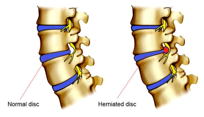
Disk herniation is a breakdown of a fibrous cartilage material (annulus fibrosus) that makes up the intervertebral disk. The annulus fibrosus surrounds a soft gel-like substance in the center of the disk called the nucleus pulposus. Pressure from the vertebrae above and below may cause the nucleus pulposus to be forced against the sides of the annulus.

The constant pressure of the nucleus against the sides of the annulus will cause the fibers of the annulus to break down. As the fibers of the annulus break down, the nucleus will push toward the outside of the annulus and cause the disk to bulge in the direction of the pressure.

This condition most frequently occurs in the lumbar region and is also commonly called a herniated nucleus pulposus, prolapsed disk, ruptured disk, or a slipped disk.


Description

The spinal column is made up of 24 vertebrae that are joined together and permit forward and backward bending, side bending, and rotation of the spine. There are seven cervical (neck), twelve thoracic (chest region), and five lumbar (low back) vertebra. There are intervertebral disks between each of the 24 vertebrae as well as a disk between the lowest lumbar vertebrae and the large bone at the base of the spine called the sacrum.

Disk herniation most commonly affects the lumbar region. However, disk herniation can also occur in the cervical spine. The incidence of cervical disk herniation is most common between the fifth and sixth cervical vertebrae. The second most common area for cervical disk herniation occurs between the sixth and seventh cervical vertebrae. Disk herniation is uncommon in the thoracic region.

The peak age for occurrence of disk herniation is between 20 and 45 years of age. Studies have shown that males are more commonly affected than females in lumbar disk herniation by a 3:2 ratio. Long periods of sitting or a bent-forward work posture may lead to an increased incidence of disk herniation.

normal and herniated disk

There are four classifications of disk pathology:
  • A protrusion occurs when a disk bulges without rupturing the annulus fibrosus.
  • A prolapse occurs when the nucleus pulposus pushes to the outermost fibers of the annulus fibrosus but does not break through them.
  • An extrusion occurs when the outermost layer of the annulus fibrosus is torn and the material of the nucleus moves into the epidural space.
  • A sequestration occurs when fragments from the annulus fibrosus or the nucleus pulposus have broken free and lie outside the confines of the disk.

Causes & symptoms

Any direct or, forceful in a vertical direction pressure on the disks can cause the disk to push its nucleus into the fibers of the annulus or into the intervertebral canal. A herniated disk may occur suddenly from lifting, twisting, or direct injury, but more often it will occur from constant compressive loads over time. There may be a single incident that causes symptoms to be felt, but very often the disk was already damaged and bulging prior to any one particular incident.

Depending on the location of the herniation, the herniated material can also press directly on nerve roots or on the spinal cord. Pressure on the nerve roots or spinal cord may cause a shock-like pain sensation down the arms if the herniation is in the cervical vertebrae or down the legs if the herniation is in the lumbar region.


In the lumbar region a herniation that presses on the nerve roots or the spinal cord may also cause weakness, numbness, or problems with bowels, bladder, or sexual function. It is unclear if a herniated disk causes pain by itself without pressing on neurological structures. It is likely that irritation of the disk or the adjacent nerve roots may cause muscle spasm and pain in the region of the disk pathology.

Diagnosis

Several radiographic tests are useful for confirming a diagnosis of disk herniation and locating the source of pain. X rays show structural changes of the lumbar spine. Myelography is a special x ray of the spine in which a dye or air is injected into the patient’s spinal canal. The patient lies strapped to a table as the table tilts in various directions and spot x rays are taken. X rays showing a narrowed dye column in the intervertebral disk area indicate possible disk herniation.

Computed tomography scan (CT scans) exhibit the details of pathology necessary to obtain consistently good treatment results. Magnetic resonance imaging (MRI) analysis of the disks can accurately detect the early stages of disk aging and degeneration. Electromyograms (EMGs) measure the electrical activity of the muscle contractions and possibly show evidence of nerve damage.

A number of physical examination procedures may be used to determine if a herniated disk is pressing on nerve root. While these tests may not identify the definitive presence of a herniated disk, they are very useful for indicating if there is pressure on a nerve root from some structure such as a herniated disk.

The straight leg raise test may be used to identify pressure on nerve roots in the lumbar region while the Spurling’s test (involving neck motion) may be used to identify compression of nerve roots in the cervical region. Compression of nerve roots in the cervical, thoracic, or lumbar regions may be apparent with the slump test.

Herniated disk cause and symptoms

Treatment

It is unclear if herniated disks cause pain themselves, or if they must press on a nerve root to cause pain. Pain may also occur with herniated disks as a result of mechanical or neurological irritation of surrounding structures such as muscles, tendons, ligaments, or joint capsules.

Therefore, many treatment strategies will be primarily focused on managing symptoms that occur in conjunction with a herniated disk. Unless a serious neurological problem exists, most symptoms of a herniated disk will resolve on their own. Yet, the interventions listed below may greatly speed the time required to resolve symptoms associated with a herniated disk.

Chiropractic manipulations are often used to treat herniated disks. There is often significant joint restriction that accompanies a herniated disk and the manipulative therapy is effective at helping to mobilize movement restrictions in the spine. Mobilizing the spine will help the patient get back to moderate activity levels sooner.

The earlier an individual can return to moderate activity levels, the quicker they can expect a resolution of their symptoms. Chiropractic manipulations are generally done with a greater frequency when a condition is in an acute stage. The frequency of treatments will be reduced as the condition improves.

Osteopathic therapy, considered by some to be an alternative treatment, may use manipulations or manual therapy techniques very similar to those of chiropractors. However, osteopathic physicians often employ more manual therapy techniques that focus on the role of the muscles and other soft tissues in producing pain sensations with herniated disks. Osteopathic physicians may also recommend use of the same medications prescribed by allopathic physicians. Some osteopaths also perform surgery for herniated disks.

Acupuncture involves the use of fine needles inserted along the pathway of the pain to move energy through the body and relieve the pain. Neurological irritation is considered to be a frequent source of pain with a herniated disk. Many believe acupuncture is particularly effective for pain management and addressing this neurological irritation. Acupuncture can also help break the cycle of pain and muscle spasm that often accompanies a herniated disk.

Massage therapists focus on muscular reactions to the herniated disk. Neurological irritation that comes with a herniated disk will often cause excessive muscle spasms in the lower back muscles. These spasms will perpetuate dysfunctional movements in the joints of the spine and may exaggerate compressive forces on the intervertebral disk.

By relaxing the muscles, massage therapists will attempt to manage the symptoms of disk herniation until proper movement can be restored. Proper movement and avoidance of aggravating postures, like sitting for long periods, will often be a great help in completely resolving the symptoms.

Allopathic treatment

Unless serious neurologic symptoms occur, herniated disks can initially be treated with pain medication. Pain medications, including anti-inflammatories, muscle relaxants, or in severe cases, narcotics, may be used if needed. Bed rest is sometimes prescribed.

However, bed rest is frequently discouraged as a treatment for herniated disks unless movement is severely painful. It has become apparent that prolonged periods of bed rest may aggravate symptoms, slow down the healing time, and cause other complications.

Epidural steroid injections have been used to decrease pain by injecting an anti-inflammatory drug, usually a corticosteroid, around the nerve root to reduce inflammation and edema (swelling). This treatment partly relieves the pressure on the nerve root as well as resolves the inflammation. Physical therapists are skilled in treating acute back pain caused by disk herniation.

The physical therapist can provide noninvasive therapies, such as ultrasound or diathermy, to project heat deep into the tissues of the back or administer manual therapy, if mobility of the spine is impaired. They may help improve posture and develop an exercise program for recovery and long-term protection. Traction can be used to decrease pressure on the disk. A lumbar support can be helpful for a herniated disk at this level as a temporary measure to reduce pain and improve posture.

Surgery may be used for conditions that do not improve with conservative treatment. There are several surgical approaches to treating a herniated disk. A number of surgical procedures may be used to remove a portion of the intervertebral disk that may be pressing on a nerve root. When a portion of the disk is removed through a surgical procedure it is called a discectomy.

Sometimes a spinal fusion will be performed after disk material has been removed. In this process a portion of bone is taken from the pelvis and placed between the bodies of the vertebrae. A spinal fusion will limit motion at that vertebral segment, but may be helpful in the event that significant disk material has been removed.

Chemonucleolysis is an alternative to surgical removal of the disk. Chymopapain, a purified enzyme derived from the papaya plant, is injected into the disk space to reduce the size of the herniated disks. The reduction in size of the disk relieves pressure on the nerve root. In 2002, Tokyo doctors produced evidence that a growth factor called vascular endothelial growth factor (VEGF) may speed up the process of injured disk resorption.

In September 2002, a noted orthopedic and spine authority named John Engelhardt became the first American to receive an artificial disk replacement (using the Bristol disk) in an operation in Switzerland. The artificial disk technology was still in clinical trials in the United States and was not expected to be approved until about 2005 or later.

Expected results

Only a small percentage of patients with unrelenting neurological involvement, leading to chronic pain of the spine, need to have a surgical procedure performed. This fact strongly suggests that many patients with herniated disks respond well to conservative treatment. Alternative therapies can play a significant role in managing the pain and discomfort for the majority of patients with a herniated disk.

In fact, magnetic resonance imaging (MRI) studies of the lumbar spine have indicated that many people without any back pain at all have herniated disks. This finding means it is unclear what role the herniated disk plays in many back pain cases. For many of these patients, proper symptom management of pain and improvement in joint motion and mobility through manual therapies will be enough to fully resolve their symptoms. For those patients who do require surgery, options are available for newer and less invasive procedures that will allow a quicker healing time.

Prevention

Proper exercises to strengthen the lower back and abdominal muscles are key in preventing excess stress and compressive forces on lumbar disks. Good posture will help prevent problems on cervical, thoracic, and lumbar disks. A good flexibility program is critical for prevention of muscle spasm that can cause an increase in compressive forces on disks at any level.

Proper lifting of heavy objects is important for all muscles and levels of the individual disks. Good posture in sitting, standing, and lying down is helpful for the spine. Losing weight, if needed, can prevent weakness and unnecessary stress on the disks caused by obesity.

Such alternative treatments as chiropractic, massage therapy, or acupuncture may play a very important role in prevention of herniated disk problems. Regular use of these approaches may help maintain proper muscular tone and reduce the cumulative effects of postural strain that may lead to the development of disk problems.